1
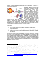
At this point let us see how to compare two or more structures. From the “Molecule File Browser” window, let us load the structure with the PDB code: 2RH1: • Click and open the “File” menu • Select “New Molecule…” entry As you have seen before, you can do it in one step by inserting such a PDB code in the “Filename” box: VMD will contact the Protein Data Bank and will download the file for you. The two structures will be visualized in the VMD OpenGL Display window but they
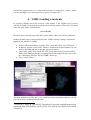
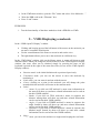
are not close each other. Sometimes, You cannot even be able to properly see the new structure just because it has a different reference system. So, we need to overlap the two proteins: • Open the menu “Extensions” in the VMD Main window, then choose “Analysis” and finally click on “MultiSeq”. • In the untitled.multiseq window tick the two structures you want to align: o 1F88_A (i.e. the chain A of 1F88 protein) o 2RH1 • Click on the button “Tool” in this window and select “Stamp Structural Alignment”: a new window will show up where some options can be choosen. • In the “Align the following” section of this new window, select “Market Structures”. All the other options can be left at their default values. • Press OK • Use “Reset view” in the Display menu of the VMD Main window to force VMD to recalculate its best view point of this new protein system. Look at the VMD OpenGL Display window: the two proteins are now “partially”
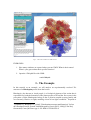
overlapped. If you change the representation to NewCartoon for both the structures
you can probably make easier your understanding about the overlapping result:11 the
alignment algorithm recognized the 7-helices common structure in the two files and
overlap them. EXERCISES
1. What are the structural differences of the 2HR1 protein with respect the 1F88
one?
2. What is the 2HR1 domain that was not overlapped to 1F88_A?
Notice that VMD has another routine for structural alignment that can be recalled by
selecting in sequence Extensions/Analysis/RSMD Calculator. It aligns by minimizing
the residue-residue mean square distance. However, this routine is rather limited
because it can align only two structures that has the same sequence and even the
11 Make sure that in the Graphical representation window the Selected Molecule be
the one just loaded and it should be referred to as molecule 1.