1
COVER STORY
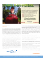
Coagulation
The complex process by which blood forms clots
Did you know that every time you bang your hand on a stair rail, or twist your ankle, your body starts forming blood clots at the injury
site? Our bodies form blood clots all the time (through a process known as hemostasis), even when we do not remember the hit or bump.
So why aren’t we one big clot? ... Because of the remarkable
biological feedback systems in our bodies. In addition to the
coagulation-promoting factors, there are also substances in
blood which inhibit coagulation. Whether or not blood coagulates
depends on the balance that exists between the two groups of
factors (pro-coagulants and anti-coagulants). When a body tissue is injured and begins to bleed, it initiates a
sequence of clotting factor activities – the coagulation cascade –
leading to the formation of a blood clot. This cascade is comprised
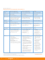
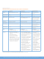
of three pathways: extrinsic, intrinsic, and common. Two laboratory tests are commonly used to evaluate coagulation
disorders: Prothrombin Time/International Normalized Ratio (PT/INR)
which measures the integrity of the extrinsic system, as well as factors
common to both systems, and Activated Partial Thromboplastin
Time (aPTT), which measures the integrity of the intrinsic system
and the common components.
When body tissue is injured and begins to bleed, it initiates a
sequence of clotting factor activities - the coagulation cascade leading to the formation of a blood clot. This cascade is comprised
of three pathways: extrinsic, intrinsic, and common.
Two laboratory tests are commonly used to evaluate coagulation
disorders: Prothrombin Time (PT/INR) which measures the integrity
of the extrinsic system as well as factors common to both systems,
and Activated Partial Thromboplastin Time (aPTT), which measures
the integrity of the intrinsic system and the common components.
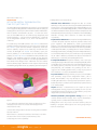
Blood Clot Formation
Clot formation is a function of plasma. It depends upon the orderly
interaction of a group of plasma proteins (which are sequentially
activated following vascular injury) with phospholipids (from either
damaged tissue or platelets) and calcium ions (Ca++). The final stages
include the formation of thrombin, which then converts soluble
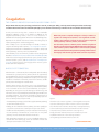
plasma protein fibrinogen to insoluble fibrin. Another factor converts the fibrin into a cross-linked polymer
which stabilizes the platelet plug and traps red blood cells (RBCs)
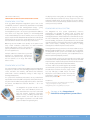
in the meshwork to form the actual blood clot. See Figure 11. Depending on the type of vascular damage or abnormality, clot
formation can be initiated and proceed according to two different
cascading pathways: the intrinsic (initiated by contact with an
abnormal/foreign surface) or the extrinsic (initiated by exposure
to tissue factors). Please keep in mind:
• The two pathways converge, so the final steps (the common pathway)
are common to both schemes.
• Clotting can be initiated via either pathway: the more rapid (15-20
secs) extrinsic scheme or the slower (2-6 mins) intrinsic scheme. • The division into two pathways is only an artifact of in vitro testing:
the two pathways interconnect at several levels. In vivo, both
pathways must be activated for effective hemostasis. • Both coagulation pathways, by a series of feedback mechanisms,
control their own activity (e.g. traces of thrombin enhance the
activity of earlier factors in the scheme). Figure 1
Thrombin is the master regulator of the coagulation cascade.
It converts fibrinogen to fibrin in order to form a loose fibrin
clot. Thrombin also activates Factor XIII, or transglutaminase,
to form stabilizing covalent bonds within the fibrin strands
to activate more platelets and form a stronger clot.
Finally, thrombin activates Factors V and VIII in a positive
feedback mechanism so the clotting cascade is
self-regulating and knows when to ‘turn off’.
>> Continued on page 6
COLA Information Resource Center 800.981.9883 | www.cola.org | www.LabUniversity.org | www.COLAcentral.com
3