1
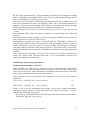
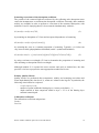
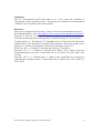
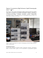
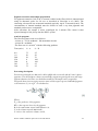
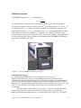
Pigment extraction and sample preparation For pigment extraction 2 ml of 90 % acetone is added to the filter which is ultrasonicated using an ultrasonic probe for 20 secs as described in Llewellyn et al. (2005). The extracting solvent also has an internal standard (typically Apo-8’-Carotenal (trans)). The concentration of internal standard must be chosen in such a way that pigments and standard peak areas are comparable. After extraction, the sample is micro centrifuged for 2 minutes The extract is then injected through a 100 l loop into the HPLC system. Analysis program The solvent systems used are as follows: - solvent A = 70:30, methanol : 1M ammonium acetate - solvent B = methanol -The flow rate is 1-ml min-1 with the following gradient: Time (min.) % A % B 0.0 1.0 20.0 25.0 30.0 30.1 39.0 75 50 30 0 0 75 75 25 50 70 100 100 25 25 Processing description Detection wavelengths are 440 nm for chlorophylls and carotenoids and 667 nm for phaeopigments. The chromQuest software automatically outputs integrated peak areas and assign pigment identities, but these are checked manually for all samples and re-assigned/reintegrated when necessary using retention times and absorption spectra. Individual pigment concentrations (Cpi) in ng L-1 are calculated as: Where: Âpi is the peak area of the pigment, RFpi is the response factor for the pigment, Bd is the ammonium acetate buffer dilution (2), Vi is the volume injected (0.0122 mL), Ve refers to the extraction volume (2 mL), IESCA satellite validation protocols 07-027-FR-ISECA 25