1
APPENDICES
TECHNICAL ARTICLES
BASIC OPERATING INSTRUCTIONS
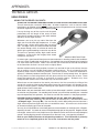
When you are looking for deep arteries, or for small or obstructed arteries, you will have to turn
the VOLUME control near maximum. This also means that every time you move the head of
the probe you are going to get some pretty big thumping noises in the earphones. Therefore
you want to avoid moving the head of the probe with respect to the skin as much as possible.
That is why you place the probe over the area where you think the artery is and then you search
for the exact point by moving the skin with the probe and changing the angle of the probe with
respect to the skin. You might wonder why these big transient noises can’t be filtered. We do
limit their intensity, but we do not filter. The reason is that in the search for low-velocity blood
flow, such as in occluded arteries and in the veins, the pitch of the Doppler sounds associated
with the blood flow are very low. Any filtering to eliminate or minimize the sounds accompanying
movement of the probe would also reduce the response to low-velocity blood flow sounds, and
of course this is undesirable.
3. DIAGNOSIS OF ARTERIAL DISEASE
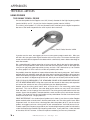
The Doppler method of diagnosing arterial disease of the limbs is only one of several good
methods. It is probably the most convenient and least expensive of the better methods. It
is only qualitative but can be made semi-quantitative by permitting you to make systolic
blood pressure measurements along the leg with the aid of a proper cuff and manometer.
The great sensitivity of the transcutaneous Doppler can cause a doctor or technician to
conclude improperly that an arterial pathway is open when it isn’t. Collateral flow around
an obstruction can be well-developed, especially in the thigh, and cause pulsatile blood to
flow in the distal arteries. Or a major artery may be narrowed, causing pulsatile flow distally.
These mistakes in diagnosis can be avoided almost entirely by simple means and a little
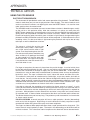
bit of experience. An experienced user of the Doppler can recognize the characteristic
sounds of open and obstructed arteries. Remember that Doppler sounds vary in pitch
(frequency) with the velocity of blood flow. When you hear the Doppler sound on a normal
artery and compare it with a normal arterial pulse-pressure wave, you will recognize the
sound of the dicrotic notch, the very fast rise time of the wave and perhaps a third sound
just before the onset of a new pulse wave. While the origin of these second and third waves
in the descending branch of a pulse wave may be in dispute, their absence in vessels distal
to an obstruction is not disputed. So a diagnostic rule is that whenever you hear the second
and perhaps third sounds of a pulse wave of a major artery, you can be sure the artery is
open proximal to the probe. Plethysmographic studies also show a delayed crest to the
wave, associated with a slower rise time to the wave when there is an obstruction proximally.
Though the Doppler is permitting you to hear velocity changes rather than true volume
changes, the correlation is good enough to be quite valuable diagnostically.
Now the opposite is not necessarily true—that when you can’t hear second and perhaps
third sounds the artery is obstructed proximally to the probe. In the digits and smaller
vessels the pulse wave is smoothed out more, especially when there is some
vasoconstriction. Now of course there are cases that are in doubt. If you cannot clearly
hear the second and third sounds (the third sound is frequently missing), compare with the
same artery on the other limb. If you find a radical difference in the sound of the Doppler,
both in pitch and in amplitude, you are justified in being quite suspicious of the patency of
the artery of the first limb you studied provided you are now fairly skilled at optimizing the
sounds.
V.2
PARKS Medical Electronics, Inc.
Aloha, Oregon
U.S.A.