1
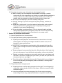
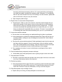
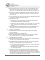
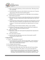
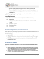
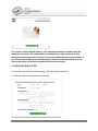
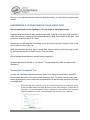
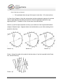
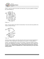
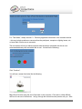
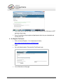
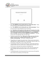
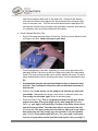
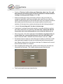
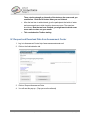
on the right. The inflated surfaces have the .sulc.native.shape.gii layer displayed.
• Rotate the surfaces around to get a good view of all aspects. Specifically look for fused
sulci, missing sections or any major additional artifacts that are clearly not cortex.
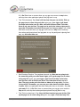
• If there are
questionable
locations, click on
them in either the
volume or the
surface views to
compare volume
vs. surface
representations for
clarification of what
might be the
problem.
HCP_<subject_id>.Q#_QC.wb.native.scene
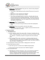
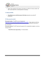
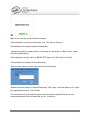
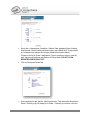
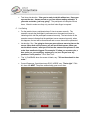
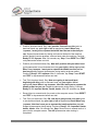
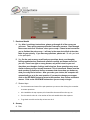
• Click on Tab 1 to get the Volume control Toolbar and scroll through the volume slices
(coronal view suggested) showing the white and pial surface outlines. Look for any errors
in the surface outline compared to the underlying volume image (anywhere the outline does
not match up the white matter boundary or the gray matter/pial surface boundary).
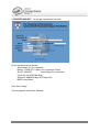
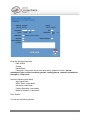
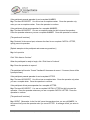
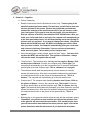
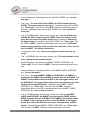
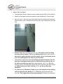
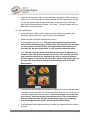
• Option: click on Tab 2 to get the Montage Toolbar and switch the surfaces to the
midthickness selection to see a more detailed view of the folding if there are questionable
regions.
(The medial wall irregularities and mismatches can be ignored. The insula and amygdala
regions tend to be messy as well; no need to report these as they are so common, unless they
are out-of-the-ordinary errors).
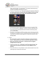
Document this QC measure in the spreadsheet titled “Overall_QC_<release>.xls”. This
spreadsheet is also located on the HCP wiki.
If everything looks good, then just put a check mark in the box for “Surface check”.
If there are any issues that may have a significant effect on further analysis, add a note in the
Comments column with the label “S-lh: or –rh:” for Surface-left hemisphere or -right hemisphere.
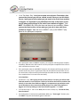
Open HCP_<subject_id>.164k_fs_LR.scene
These scenes are used for reviewing the quality of the areal distortion, myelin maps, and
volume distortion.
500 Subjects + MEG2 Reference Manual – Appendix IV | WU-Minn Consortium of the NIH Human Connectome Project Page 23