1
PRINCIPLES OF OPERATION
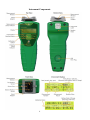
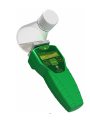
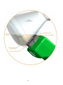
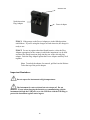
When the OD Scanner is pressed against the side of a liquid culture vessel and the
two Position Sensors on the front face of the instrument are activated, an infrared
laser behind the Measurement Window is directed into the liquid culture. The
laser light is scattered by the cells or microorganisms, creating a “glow ball” of
monochromatic light. The intensity and size of the glow ball is dependent on the
biomass within the liquid culture. At early stages of growth, when the biomass is
low, the glow ball will be large in size and weak in intensity. As the cells or
microorganisms grow and divide, the concentration will increase and the glow
ball will reduce in size and increase in intensity. The detectors behind the
Measurement Window, surrounding the laser, are sensitive to the intensity of light
back-scattered from within the glow balls. The measured intensity is combined in
a manner that is linear with biomass over a very wide range of response.
Although classical Optical Density (OD) measurements in a spectrophotometer
require dilution in order to accurately determine biomass greater than about 0.5
g/L, the OD Scanner is able to determine biomass from 0.05 to 10 g/L or more,
without dilution, or any liquid handling.
One of the advantages of using an infrared laser source for measuring biomass is
the avoidance of light absorbance by colored media components (and colored
vessel materials). This allows for the measurement of true scattering rather than a
combination of absorbance and scattering. As a result, a highly linear relationship
is maintained between biomass concentration and the measured optical
reflectance. However, it is also important to realize that for biomass containing
strongly visible-light-absorbing chromophores (e.g. photosynthetic algae), the
chromophore absorbance may affect the agreement between a conventional OD
measurement and the result reported by the OD Scanner. The OD measured in
the visible range by a spectrophotometer will be influenced by both chromophore
absorbance and cell scattering, whereas the OD reported by the OD Scanner will
be based only on cell scattering. In such situations, if the relationship between
chromophore concentration and biomass is relatively fixed, it still may be possible
to generate a strong correlation between OD measured by conventional methods
and that reported by the OD Scanner. However, it is important to be aware that
changes in chromophore concentration that are not accompanied by biomass
change would not affect the result reported by the OD Scanner, but would skew
the results determined by conventional visible spectrophotometry.
Another important consideration when comparing OD Scanner results with
conventional spectrophotometry, is the optical design of the spectrophotometer.
Most spectrophotometers are designed to make accurate measurements of
absorbance, but not scattering. A determination of chromophore absorbance
requires only a comparison of how much light is extinguished within a sample
when the chromophore is present at different concentration levels (e.g. zero and a
known concentration). By contrast, in a scattering measurement, light is deviated
from its path, rather than being extinguished. As a result, the measured amount of
scattering will be dependent on the area and angle of scattered light that is
captured by the detection system. Since the detector size and geometric
arrangement is not standardized between different commercial
19